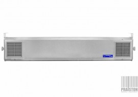


- Recodnitioned (used),
- Technical condition: very good,
- Visual condition: very good,
- Real photos of the product,
- Made in Japan (2015),
- Power supply: 230 V,
- Frequency: 50/60 Hz,
- Power: 250 VA,
- Swept Source Technology (SS OCT) offers new possibilities for imaging the structures of the eye. Compared to the most widely used spectral imaging devices (SOCT), it enables gaining much faster scanning speed, much better penetration of the eye's structures up to the sclera, and better final resolution of the obtained images. The use of 1050nm (infrared) wavelength in SS OCT TRITON provides much better tissue penetration than the red light used in SOCT. The infrared light also undergoes less scattering, allowing better visualization of the posterior vitreous cortex and its structure. Thanks to the swept source properties described above, the DRI OCT TRITON is an instrument that allows to look deeper into the structures of the human eye. Extensive glaucoma functions with a normative database are implemented in the device. Thanks to the large scanning area, it is possible to make an imaging and qualitative diagnosis of the macular area in one scan, measure the thickness of the nerve fiber layer around the disc, ganglion cells in the central area, and obtain topographic data of the optic nerve disc,
- Main advantages:
- simultaneous high-quality images of the vitreous body, retina, choroid and sclera,
- visualization of the vitreous with the ability to perceive the differentiation of its structure,
- automatic mapping of retinal and choroid thickness and volume layers,
- obtaining much better quality of OCT scans in patients with reduced translucency of optical centers, including cataracts and harmful refractive error,
- thanks to its large maximum scan width (12mm), the device has the ability to image the macular area and the nerve disc simultaneously,
- high transmittance and low light scatter allow for high-quality, artifact-free anterior segment cross-sections,
- lack of scan lines visible to the patient, which often cause loss of fixation and deterioration of the examination quality,
- Specification:
- The fundus of the eye photography and observation
- Type of photography: Color, non-infrared,
- Photography angle: 450, 300 equivalent (digital zoom),
- Operating distance: 34.8 mm,
- Minimum pupil’s diameter:
- Normal: 4.0 mm or more,
- Narrow: 3.3 mm or more,
- Fundus tomogram observation and photography
- Scanning range (eye fundus):
- Horizontal: 3 do 12mm,
- Vertical: 3 do 12mm,
- Scanning profiles:
- 3D scan,
- Line scan (linear, cross-type, radial),
- Scanning speed: 100 000 A-scans per second,
- Horizontal resolution: 20 µm,
- Axial resolution:
- Digital: 2.6 µm,
- Optical function: 8 µm,
- Minimum pupil’s diameter: 2.5 mm or more,
- Scanning range (eye fundus):
- Fundus eye observation and photography / fundus tomogram
- Fixation point::
- Internal fixation point:
- Organic EL point matrix,
- Display position can be changed and adjusted,
- Display method can be changed,,
- Peripheral fixation point:
- It is displayed according to the position of the internal fixation point,
- External fixation point,
- Internal fixation point:
- Fixation point::
- Frontal observation and photography
- Photography type: IR,
- Operating distance: 17 mm,
- Observation and photography of the anterior segment tomogram
- Operating distance: 17 mm,
- Scanning range (on cornea):
- Horizontal: 3 do 16mm,
- Vertical: 3 do 16mm,
- Scanning profiles:
- 3D scan,
- Line scan (linear, radial),
- Scanning speed: 100 000 A-scans per second,
- Fixation point:
- Internal fixation point,
- External fixation point,
- TOPCON IMAGEnet6 ver. 1.03 (1.35.20386) software, in English with lifetime licence,
- English keyboard on the control panel,
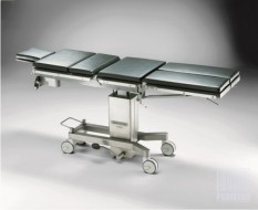
- The whole unit is placed on OPTOPOL SE-275 mobile table (has traces of use) with electric height regulation in the range of: 64-84 cm,
- In the set:
- Instruction manual in the following languages: PL, EN,
- PC computer with WIN8 EN + keyboard and a mouse,
- LG 23” IPS 23MB35PYI monitor,
- HP Color LaserJet CP1215 printer,
- TOPCON Kit AA-1 Anterior Eye Examination Attachment,
- Fixation point,
- Case,
- Set of power cords,
- Dimensions:
- OCT: 50 x 28 x 60 cm,
- Table top: 120 x 52 cm,
- Overall: 52 x 140 x 140 cm,
- Weight:
- Of the table: 72 kg,
- OCT: 22 kg,
- Overall: 135 kg,
- Provided with a valid Technical Passport,
- Guarantee:
- 6 months for the domestic market (Poland),
- 3 months for the international market,
- Financing possibilities (Poland only): Installments, leasing, loan,
Reconditioned


Recently Viewed Items
KARL STORZ Surgical Instrument Set
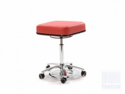
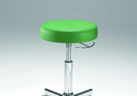
Medical Stool Coburg Medicalift 3030
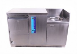
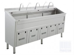
MEIKO TOPLINE 40 Duck and pool washer