

Reconditioned (used),
Technical condition: very good,
Visual condition: very good,
Real photos of the product,
Japanese production,
Power supply: 230 V,
Frequency: 50/60 Hz,
Power: 900 VA,
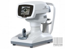
HITACHI
Aloka F37
is a compact ultrasound machine that has very good image quality with volumetric ultrasound beam processing technology. It is a new imaging quality with very fast and precise imaging. The Aloka F37 has a very wide operating range,
Camera features:
Broadband Harmonic (BbH) - broadband harmonic imaging - provides crystal clear images with elimination of most common artifacts, but without sacrificing high penetration of the object under examination,
Silky Image Processing (SIP) - advanced smoothing image processing - a software-implemented function effectively removes artifacts and noise that interfere with diagnosis and accurately highlights the difference between tissue boundaries. Optimal processing can be automatically configured according to each clinical application,
Adaptive Image Processing (AIP) - thanks to hardware background processing, it does not slow down the image in the slightest, while allowing much better extraction of tissue contrasts and contours of imaged structures,
Spatial Compound Imaging (SCI) - compound imaging (multi-angle scanning) significantly reduces random noise, while eliminating image artifacts resulting from anisotropic reflection of ultrasound at different angles,
Extended Flow (eFlow) - a Doppler imaging mode with increased temporal and spatial resolution, in addition to the excellent conventional and power Doppler also available in the system, allowing better distinction of true vessel outlines and precise visualization of even very fine flows,
Real time 3D (4D) - real time 3D imaging so-called 4D. Thanks to advanced technology developed by Japanese engineers at Hitachi Aloka, 4D mode enables extremely fast and realistic real-time 3D images,
4Dshading - innovative fetal rendering technology (real-time 3D image) with natural shading and skin texture visible under incident light. A virtual light source with infinite adjustment can be placed in any position, allowing detailed observation of the smallest details of the examined fetus,
Needle Emphasis (NE) - the function allows transmission of the ultrasound beam perpendicular to the biopsy needle (independent of B-mode imaging) for better visualization of the needle,
Free Angular M-mode (FAM) - a unique anatomical M-mode with the ability to reconstruct M-mode recordings in any image section (real-time or Cine loop-based), superbly facilitating echocardiography in children or difficult patients and fetal echocardiography,
Pulsed Doppler (PW Doppler / PW HPRF Doppler) and excellent Continuous Doppler (CWD),
Color/Power Doppler - new generation dynamic wide-band Color/Power Doppler modes provide accurate analysis of blood flow morphology with excellent sensitivity and exceptional quality,
Tissue Doppler Imaging (TDI) - Color and Spectral Tissue Doppler,
Dual Dynamic Display (DDD) - an option for parallel display of the superimposed flow image next to the black-and-white image, very helpful for vascular and cardiovascular studies,
Dynamic Slow-motion Display (DSD) - dynamic slow-motion view - simultaneous display of real-time B-mode image and slow-motion B-mode image with adjustable playback speed,
Specially developed zoom mode (Zoom) without loss of temporal and spatial resolution,
Trapezoidal and rhomboid imaging for structures beyond the normal field of view of the linear head,
Built-in Image Archiving System - a fast, intuitive, easy-to-use system for archiving and processing ultrasound images and film sequences together with a patient database, reports and comments, allowing to store images on hard disk (HD), flash memory of Pen-Drive type (not included) with the possibility of data export and transmission to a computer network compatible with DICOM 3.0,
The device works with heads:
CONVEX for abdominal examinations,
LINEAR for vessels and superficial organs (including very high frequency - up to 18 MHz),
SECTOR PHASED-ARRAY for transthoracic and transesophageal echocardiography,
ENDOKAWITARY for vaginal and rectal examinations,
4D OBJECTIVE (Volume Probe),
BIOPSY with a central canal guided through the head with various types of MEDIOPERATIVE and LAPAROSCOPE heads,
Number of active ports for connecting imaging heads: 3,
Mobile on casters with brake,
Menu in language: English,
Keypad on control panel in language: English,
Included:
Convex head UST-9123,
Linear head UST-5413,
Sector head UST-5299,
Sector head UST-5298
Power cord,
Dimensions: 64 x 105 x 164 cm,
Weight: 75 KG,
It has a current inspection and is ready for operation,
Issued Technical Passport (Service Report) valid for 12 months,
Warranty:
6-months for domestic market (Poland),
3-months for the international market,
Possibility to extend the warranty up to 12-months for domestic and international market for a surcharge,
Financing options (Poland only): Installments, Leasing, Loan,


Recently Viewed Items
Tested disposable breathalyser TEST&DRIVE Sober Morning. 1 pc

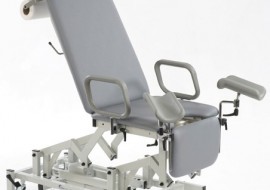
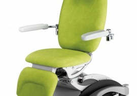
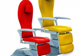
Examination Chair TEYCO MED FRANCY B3
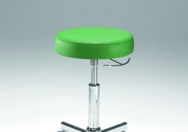
Medical Stool Coburg Eurolift 11070